Clinical Considerations in Management of Complex Apical Root Canal Anatomies
Root canal is a complex three dimensional system within tooth. Wide range of complex variations exist in root canal anatomy. It is made further complex with the presence of lateral ramifications, extra root or extra canals. Apical third of the root is always with highest percentage of ramifications and accessory canals. Among all the teeth molars and premolars present with the highest incidence of aberrant morphology.
Lateral canals are accessory canals extending from main canals to periodontal ligament space. Accessory canals contain connective tissues and vessels but do not supply the pulp with collateral circulation . Lateral canals or accessory canals can be observed anywhere along the root length but more commonly in apical third. Study by Dr Stephen Cohen concluded that 73.5% accessory canals are in apical third , 11.4% in middle third and 15.1 % In cervical third. Lateral canal is located at right angle to main root canal while Accessory canal branches off from main root canal in apical region. Similarly furcation canal are seen at furcation area of multi-rooted tooth. Apical Delta refers to system of primary or secondary canals that terminates short of the apex with lateral canals fanning out from this point to the end of root surface
Ramifications and accessory canals were increasingly eliminated by
1mm root end resection-52%
2mm root end resection-78%
3mm root end resection- 98%
This is the reason why during apicoectomy 3mm apical root removal is recommended.
The overall success of endodontic treatment depends on whether all canals are accessed, cleaned, shaped and filled .
Scouting the canal system with hand files helps in locating minute canals but it’s the Irrigation and activation protocol that holds the key.
I suggest these key points that can help in filling maximum apical anatomies.
1) Practice scouting for exploring anatomy in apical third of canal.
2) Always confirm whether irrigant is reaching in apical third of canal in full strength and concentration.
3) Activate both Sodium Hypochloride and liquid EDTA alternatively.
4) Increase time and cycles of activation so as to clean maximum lateral or accessory canals.
5) Warm vertical compaction technique of obturation is the best to fill all complexities in root canal system due to hydraulic pressure created while doing vertical compaction
6) Take time and do one canal at a time while obturation.
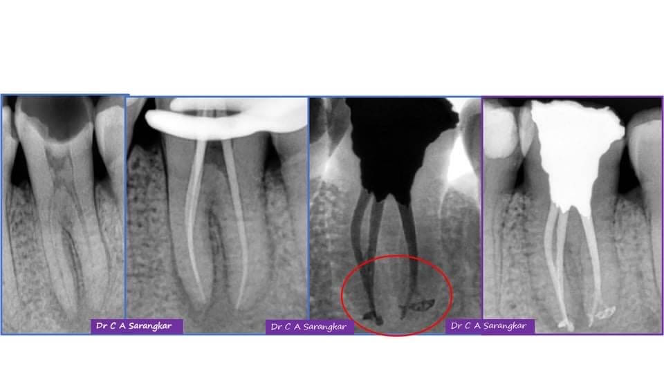
(Case done using C Pilot hand files (Dentsply), NT Gold rotary files (Nineten),EQ-S Irrigation Device (Meta Biomed), Non Standardised GP Points (Sure Endo) ,Ceraseal bio ceramic sealer (Meta Biomed)
AUTHOR

Very nice,well explained
Very well explained